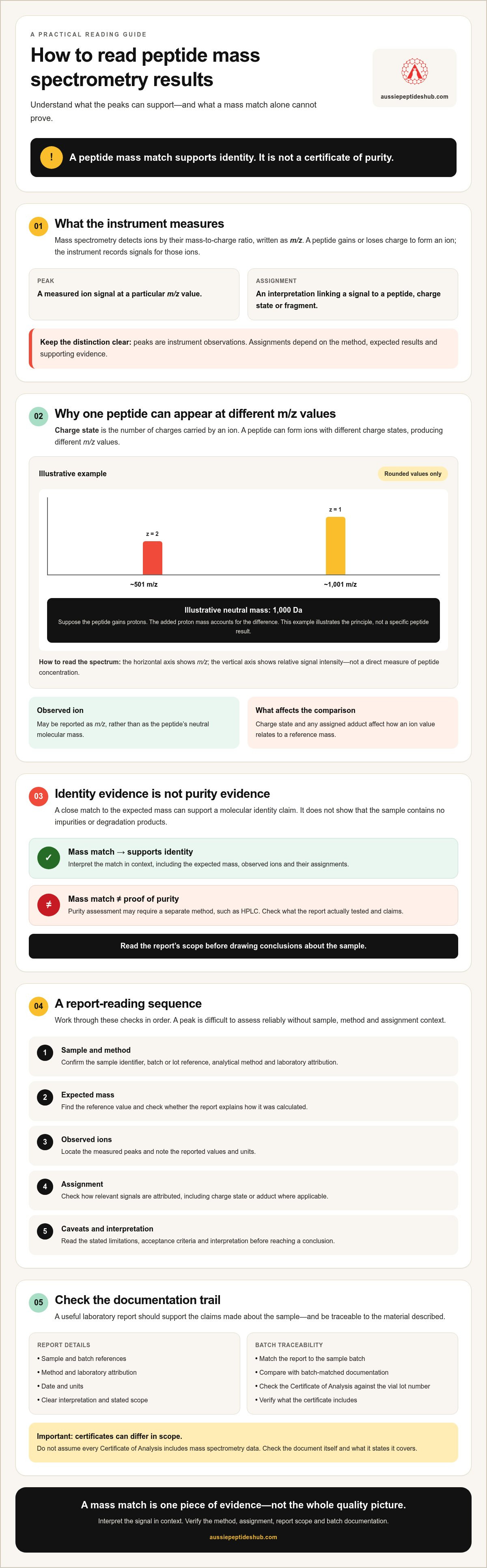
Understanding Peptide Mass Spectrometry Results: A Practical Guide
October 1, 2026 · 16 min read
A peptide mass match is evidence of identity, not a certificate of purity. If a report lists unfamiliar m/z values, charge states and mass error figures, understanding peptide mass spectrometry results starts with knowing what each measurement supports and what it leaves unresolved. A close match to the expected mass can support a molecular identity claim, but it doesn’t show that the sample contains no impurities or degradation products.
A useful laboratory report should support the claims made about a sample. To assess it, separate evidence of identity from evidence of purity, then check whether the method and documentation support each conclusion. This guide explains how to read key fields in a spectrum or report, including observed mass, charge state and mass error. It also covers why purity assessment may require a separate method, such as HPLC, and what a single result cannot establish.
You’ll also learn what supporting information to verify, including whether the report is linked to the sample’s batch. For research-grade products, a matching Certificate of Analysis and vial lot number can help with traceability. However, don’t assume every certificate includes mass spectrometry data. Check the document and its stated scope.
Key Takeaways
- When understanding peptide mass spectrometry results, check the sample details and method before interpreting peaks.
- Consider how charge states, adducts and instrument conditions can affect observed ions and their assignments.
- Treat a mass match as evidence that supports identity, not as a stand-alone measure of purity.
- Check the report for sample and batch references, method, date, laboratory attribution, units and a clear interpretation.
- Compare the report’s scope with batch-matched documentation, including the Certificate of Analysis, before drawing conclusions about a sample.
What peptide mass spectrometry measures, and what the result represents
Mass spectrometry measures ions by their mass-to-charge ratio, written as m/z. A peptide is not usually measured as a neutral molecule. It first gains or loses charge to form an ion. The instrument records signals for those ions, and analysts use the measured values and method context to assess molecular mass. With suitable fragmentation and analysis, the results can also support identification of structural features.
A measured signal is an instrument observation. The identity assigned to that signal is an interpretation that depends on the method and supporting evidence.
What does m/z mean in a peptide spectrum?
m/z means an ion’s mass divided by its charge. It isn’t automatically the peptide’s molecular mass. The charge state is the number of charges carried by an ion. A single peptide can therefore appear at more than one m/z value if it forms ions with different charge states.
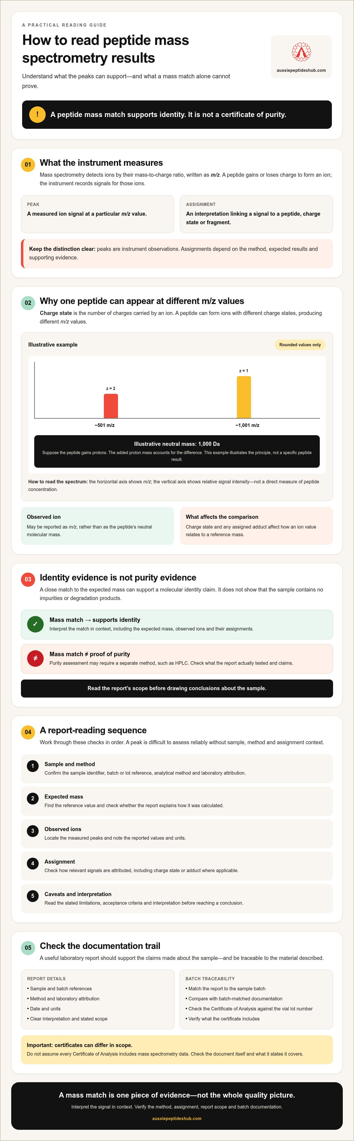
Illustrative example: Suppose a peptide has a neutral mass of 1,000 Da and gains protons. As a singly charged ion, its approximate m/z is 1,001. As a doubly charged ion, its approximate m/z is 501. The added proton mass accounts for the difference. These rounded values illustrate the principle, not a specific peptide result.
Schematic spectrum, illustrative only
Y-axis: relative signal intensity
│ │ │
└──────┬────────┬────── X-axis: m/z
~501 ~1,001
z = 2 z = 1
The horizontal axis shows m/z, with each peak marking a detected ion signal at a particular value. The vertical axis shows signal intensity, not a direct measure of peptide concentration. For a broader overview of protein analysis approaches and ionisation techniques, see Protein mass spectrometry.
How does mass spectrometry detect peptide ions?
The sample is ionised, producing charged particles that the instrument can detect. Electrospray ionisation is a common approach for peptides, but it isn’t universal. Laboratories select methods and instrument configurations to suit the sample and analytical purpose. The instrument separates ions according to m/z and records their signals. In suitable workflows, ions can also be fragmented, and the resulting pattern may support structural identification.
When understanding peptide mass spectrometry results, keep the distinction clear: peaks are measured signals. Assigning a peak to a peptide, charge state or fragment requires interpretation in light of the method and expected results.
How to interpret peptide MS results: read the spectrum in sequence
Read the report in a fixed order so you don’t interpret a peak before confirming what sample was analysed and what the method measured. When understanding peptide mass spectrometry results, use these checks:
- Sample and method: Confirm the sample identifier, batch or lot reference, analytical method and laboratory attribution.
- Expected mass: Find the reference value and check whether the report explains how it was calculated.
- Observed ions: Locate the measured peaks and note their reported values and units.
- Assignment: Check how the analyst attributes each relevant signal, including charge state or adduct where applicable.
- Caveats: Read the stated limitations, acceptance criteria and interpretation before drawing a conclusion.
Follow the checks in order. An unexplained peak can’t be assessed reliably without knowing the sample, method and assignment.
Expected mass versus observed mass
The expected mass is the reference value for the peptide under the stated calculation or reporting convention. The observed value comes from the detected ion signal. These values may not be directly interchangeable: a spectrum may report an ion’s m/z, while the reference is a neutral molecular mass. The charge state and any assigned adduct affect the relationship between them.
Compare expected and observed mass on a like-for-like basis, using the same mass convention and accounting for the reported ion assignment.
If the expected value is missing, ask which reference was used. If a peak has no clear assignment, request clarification rather than assuming it represents the target peptide or an impurity.
Charge states, adducts and mass error
A charge state is the number of charges carried by an ion. An adduct is an additional atom or molecule associated with the ion, which can shift its measured m/z. Either may produce extra peaks without indicating a different peptide or a contaminant. Instrument settings and analytical conditions can also affect the observed signal and its assignment.
Mass error describes the difference between an observed value and its expected reference. Reports may express it in daltons or parts per million (ppm). Check the units, the values being compared and the calculation or method context. There is no universal acceptable limit: the relevant criterion depends on the validated method and the laboratory’s stated requirements. Don’t apply a threshold from another report without confirming that it is relevant.
When reviewing research supplier records, check that batch references align across documents. Aussie Peptides states that products are independently HPLC-tested and supplied with matching Certificates of Analysis. HPLC testing is distinct from mass spectrometry. You can review research peptide documentation and check what a specific certificate reports before relying on it.
Does peptide mass spectrometry prove purity? Identity and purity are different
A mass match in mass spectrometry (MS) can support a peptide identity assignment, but it doesn’t, by itself, establish sample purity. Identity asks whether measured evidence is consistent with the target molecule. Purity asks what else is present and in what proportion. These are separate questions, and each requires evidence suited to it.
A single analytical result cannot verify every aspect of a sample.
What MS can support, and what it cannot establish alone
An intact-mass result can support an assignment of molecular mass. It doesn’t necessarily establish the complete structure or sequence. Tandem MS can fragment ions and produce sequence-related evidence when the method and interpretation are suitable. The strength of that evidence depends on the data and analytical approach, not simply on whether a matching mass appears.
MS signal intensity also isn’t a direct measure of each component’s proportion in a sample. Co-eluting compounds may be difficult to distinguish, and components that ionise poorly may produce weaker signals than their actual amount would suggest. A clean-looking spectrum therefore doesn’t necessarily mean the sample is free of other components.
How HPLC results answer a different question
High-performance liquid chromatography (HPLC) separates components as they pass through a column and can produce a chromatographic profile. The profile can show detected peaks and their relative areas under the stated conditions. However, peak area depends on the method, detector response and reporting approach. It isn’t automatically an absolute measure of mass purity, and components that aren’t separated or detected may not appear as distinct peaks.
MS
Question answered: Is the measured ion evidence consistent with an expected molecular mass or, with suitable fragmentation, structural features?
Typical output: Mass-to-charge signals and assigned ions or fragments.
Limitation: A mass match alone doesn’t quantify all sample components or establish purity.
HPLC
Question answered: What components are separated and detected under the method conditions?
Typical output: A chromatographic profile with retention times and reported peak areas.
Limitation: Peak area is method-dependent and doesn’t automatically equal absolute mass purity.
Purity interpretation depends on the validated method, its separation and detection capabilities, and the report’s stated calculations and criteria. Read those details before treating a percentage as a complete description of the sample. For more on chromatographic reporting, consult a dedicated guide to HPLC-tested peptides. The method’s specific conditions still matter.
For research materials, keep each document within its stated scope. Aussie Peptides states that its products are independently HPLC-tested and supplied with matching Certificates of Analysis. That information should not be taken to mean mass spectrometry was performed or that one test proves the other. Understanding peptide mass spectrometry results means evaluating the evidence from each method separately.
A practical checklist for reviewing a peptide mass spectrometry report
A report is useful only when its findings can be connected to the sample and interpreted within the method’s stated scope. Use this checklist before relying on its conclusions. For understanding peptide mass spectrometry results, traceability and clear assignments matter as much as the headline result.
Check sample identity and report traceability
- Match the sample: Confirm the name or identifier on the report matches the material being evaluated. Resolve differences in spelling, codes or sample descriptions before comparing results.
- Check the batch or lot reference: Look for a batch or lot number that links the report to the relevant sample. Without a clear sample link, the report may not establish that the result applies to the material in hand.
- Read the dates accurately: Distinguish the test date from the report issue date. Treat dispatch information as a separate record, not evidence of when testing occurred.
- Identify the laboratory: Check that the report names the laboratory or otherwise makes the source of the result clear.
Assess method, assignments and limitations
- Find the method: Check which analytical method was used and whether the report gives enough context to understand what was measured. Don’t assume a Certificate of Analysis includes mass spectrometry data.
- Check expected and observed values: Confirm the expected mass, observed signals, units and ion assignments are stated. Make sure the values are compared on the same basis, accounting for charge state or an assigned adduct where relevant.
- Review the interpretation: Look for an explanation of how the reported peaks support the stated conclusion. An unlabeled peak is not self-explanatory.
- Note limitations and discrepancies: Record missing reference values, ambiguous labels, unexplained deviations or unclear acceptance criteria. Ask the report issuer to clarify these points rather than inferring identity or purity.
Check that the document is internally consistent. For example, a reported assignment should correspond to the stated observed value and units. If the interpretation makes a broader claim than the described method supports, request method details or supporting records before accepting it.
For research-grade products, a matching Certificate of Analysis and a vial lot-number check can help connect documentation to a batch. Aussie Peptides states that its products are supplied with matching Certificates of Analysis and that lot numbers enable online batch-detail checks. Verify the specific document and its contents. Don’t assume every certificate reports a mass spectrometry result.
Use the checklist to assess the evidence without extending its scope. Review research peptide documentation and check what each batch record actually reports.
Use MS results alongside batch-matched documentation
Mass spectrometry (MS), high-performance liquid chromatography (HPLC) and a Certificate of Analysis (COA) provide different forms of analytical context. MS may support a molecular-mass assignment or, with a suitable method, provide structural evidence. HPLC separates detected components and reports a chromatographic profile. A COA brings stated test information together for a product or batch. Review each record to see what it actually documents.
No single document proves more than its method, sample and stated scope allow. A mass match doesn’t establish purity. An HPLC profile doesn’t automatically confirm complete molecular structure. A COA isn’t evidence of a test or result unless it identifies that information. Understanding peptide mass spectrometry results means considering these records together without treating them as interchangeable.
What a Certificate of Analysis can add
Match the COA to the relevant product and batch or lot. Compare the identifiers on the certificate with the material’s label and any associated report. Then review the methods and results listed. Don’t assume a COA includes MS because it is an analytical document, or that an HPLC result confirms an MS finding.
A BPC-157 sourcing and verification guide can be a useful example of how to assess batch documentation. Apply the same checks to any product record: confirm its reference to the material, examine the stated test information and note what remains outside the document’s scope.
Questions to ask before relying on a result
- Which sample was tested? Check its name or identifier and whether it matches the material being evaluated.
- Which method was used? Confirm whether the result comes from MS, HPLC or another stated method, and what analytical question it addresses.
- Is the report batch-specific? Check whether its batch or lot identifier matches the supplied material. If identifiers differ or are absent, ask how the report is linked to the sample.
- What does the result support? Compare the conclusion with the method and reported evidence. Request clarification if the claim extends beyond what’s documented.
Aussie Peptides states that its research-grade products are independently HPLC-tested, supplied with matching COAs and traceable through online lot-number checks. These details support document traceability. They don’t mean MS is performed on every product or included in every COA. Verify the current batch record and its stated contents. Products are for laboratory research use only and are not for human consumption.
For a practical next step, check available research peptides and batch documentation, then review the records for the specific material and lot.
Make each result answer the right question
Understanding peptide mass spectrometry results means distinguishing a measured mass from the interpretation assigned to it. A mass match can support an identity assessment, but it doesn’t establish purity on its own. Read the method, expected mass, observed ions and assignments together, then consider what the report leaves unresolved.
Use MS and HPLC as distinct sources of evidence, and check that documents refer to the relevant sample and batch. A Certificate of Analysis supports conclusions only within the methods and results it states.
Aussie Peptides supplies research-grade products for laboratory use only, not for human consumption. Products are independently HPLC-tested and supplied with matching Certificates of Analysis. Vial lot numbers can be used for online batch-detail checks. These records support traceability; they don’t mean that MS is performed on every product or included in every COA.
Review research peptides and batch documentation and check the records for the specific material you’re assessing. Clear identifiers and method-specific evidence make it easier to evaluate each claim carefully.
Frequently Asked Questions
What does a peptide mass spectrometry result tell you?
A peptide mass spectrometry result reports signals from ions, usually as mass-to-charge (m/z) values, and may support an expected molecular-mass assignment. With suitable methods, including fragmentation analysis, it can also provide structural or sequence-related evidence. Interpretation depends on the sample, method, charge states, adduct assignments and stated limitations. A matching mass supports an assignment; it doesn’t establish purity or verify every claim made about the sample.
Does mass spectrometry measure peptide purity?
Mass spectrometry doesn’t automatically measure peptide purity. It detects ions, but different sample components can ionise with different efficiencies, so signal intensity may not represent their proportions. Some components may also be difficult to distinguish or detect. HPLC provides a separate chromatographic profile, but its peak areas are method-dependent too. To assess purity, review the method, separation, detection and reporting details rather than treating one MS signal as a purity percentage.
How do you read m/z on a peptide mass spectrum?
Read m/z as the mass of an ion divided by its charge, not automatically as the peptide’s neutral molecular mass. In a typical spectrum, the horizontal axis shows m/z and the vertical axis shows signal intensity. Peaks mark detected ions. A peptide may produce several peaks because it carries different charge states or forms adducts. Check the report’s assignments before deciding which peak corresponds to the expected peptide.
What is the difference between MS and MS/MS for peptides?
In a basic MS measurement, the instrument records ions and their m/z values. In tandem mass spectrometry, written MS/MS, a selected ion is fragmented and the resulting product ions are measured. Those fragments can provide additional structural or sequence-related evidence. MS/MS therefore offers information beyond an intact-mass measurement, but its usefulness depends on the method, the fragments observed and how the data are interpreted.
Can a mass spectrometry result confirm a peptide sequence?
A mass match alone generally supports a molecular-mass assignment, not complete sequence confirmation. Suitable MS/MS analysis can generate fragment patterns that provide sequence-related evidence. Review the report to see which fragments were detected, how they were assigned and whether the interpretation addresses the sequence in question. The method and available data determine the strength of the evidence; don’t treat an unexplained mass peak as proof of a full sequence.
What should a peptide mass spectrometry report include?
A useful report should identify the sample and, where applicable, its batch or lot reference. Check for the method, test or report date, laboratory attribution, expected mass, observed values, units, ion or fragment assignments and an interpretation. It should also make relevant limitations clear. Confirm that the identifiers connect the result to the material being assessed, and ask for clarification if key values or assignments are missing.
Is a matching mass enough to prove a peptide sample is high quality?
No. A matching mass can support identity, but it doesn’t establish purity, quantify every component or verify all aspects of sample quality. When understanding peptide mass spectrometry results, consider the method’s scope and compare it with batch-matched documentation and other relevant analytical evidence, such as HPLC results. Check that the records identify the sample or lot and don’t claim more than the reported methods establish.